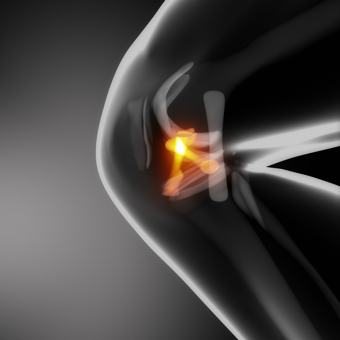
Painful patella (chondromalacia patellae) has numerous causes.
The cause of retropatellar cartilage damage is a mismatch between load and load-bearing capacity of the femoropatellar joint.
The patellotrochlear contact pressure increases due to defects in the shape and position of the patella.
Often, overloading of the corresponding joint is responsible for the complaints. Sports or occupational activities can stress the joint and damage the cartilage substance.
Accidents or falls are also among the causes of cartilage softening or its damage.
Numerous congenital anatomical reasons are decisive for this clinical picture.
If the load-bearing limit of the hyaline cartilage is exceeded, softening, fracturing and finally cartilage loss occur.

The localization of the pain in the anterior section of the knee joint is indicative, with pain intensified by walking uphill or down stairs, after prolonged sitting, or during the transition from a physically less active lifestyle to high stress, such as the start of a vacation.
Clinically, chondromalacia can be identified by increased retropatellar crepitation; an effusion may also form due to abrasion. Furthermore, a patella pressure pain can be triggered during the clinical examination, which is intensified by tensing of the quadriceps femoris muscle with passively fixed patella (sole sign).
Lateral radiographs may show increased subchondral sclerosis; tangential radiographs at 30°, 60°, and 90° flexion assess whether the patella tilts or lateralizes (malalignment).
Cartilage can be visualized on magnetic resonance imaging; however, cartilage quality is far more assessable by arthroscopy. Cartilage damage can be classified arthroscopically; the ICRS classifications have become established.
Differentially, all other causes of femoropatellar pain syndrome can be considered. Frequently, there are mechanical overloads of the structures inserting at the patella, especially the quadriceps and patellar tendons (see patellar tendon syndrome). Furthermore, bursitis of the bursa pre- or infrapatellaris may cause similar symptoms.
The correct therapy for the patient can usually be initiated subsequently.
If weak muscles are the cause of patellar instability, physical therapy can make the symptoms disappear within as little as one to two months. The condition of the extensor muscles on the thigh in particular, especially the middle and inner muscle belly, plays an important role in stabilizing the patella in its normal gliding path. Strengthening the knee extensors is therefore given special emphasis in a 4-phase program. In extensive mechanical studies, the direction of pull of the individual thigh extensor muscles was precisely determined and the influence of the individual muscle pulls on the sliding path of the kneecap was investigated. If the muscle portion located toward the inner side of the thigh is too weak, it cannot perform its important function, which is to prevent the patella from shifting and tilting outward during extension.
The muscle weakness may cause occasional outward dislocation of the kneecap. Therefore, in order for the kneecap to slide normally again, it is imperative to strengthen this muscle.
Dear Patients,
Please note our special opening hours in April and May:
Easter holidays:
17.04.25 Practice Datteln closed
23.04.25 Practice Recklinghausen and Datteln closed
24.04.25 Practice Recklinghausen and Datteln closed
25.04.25 Practice Recklinghausen and Dortmund closed
May:
02.05.25 Practice Kamen closed
23.05.25 All practices incl. telephone exchange closed
